
Tissues and Supporting Systems in Animals – SS2 Biology Lesson Note (First Term)
Lesson note at a glance
- Class
- SS2
- Subject
- Biology
- Term
- First Term
- Reading time
- 12 min
A skeleton is the framework of the body that supports an animal, gives it shape and protects its soft tissues and organs.
Behavioural objectives
By the end of the lesson, students should be able to:
- name the three forms of skeletal materials and state where each is found;
- state the differences between bone and cartilage;
- describe the hydrostatic skeleton, exoskeleton and endoskeleton;
- list the functions of the skeleton;
- name the bones of the axial and appendicular skeletons;
- describe the features and functions of the different types of vertebrae.
What are tissues?
A tissue is a group of similar cells that work together to carry out a specific function.
- Epithelial tissue: covers body surfaces and lines organs, e.g. the skin and the lining of the gut.
- Connective tissue: supports and joins other tissues, e.g. blood, bone and cartilage.
- Muscular tissue: contracts to bring about movement.
- Nervous tissue: carries impulses between parts of the body.
What is a skeleton?
The skeleton is the framework of the body that gives support, shape and protection to the soft tissues and organs of an animal.
- Materials: it is made of chitin, cartilage or bone.
- Kinds: it may be a hydrostatic skeleton, an exoskeleton or an endoskeleton.
- Parts in vertebrates: the axial skeleton and the appendicular skeleton.
- In man: it forms the central core of the body and is covered by muscles, blood vessels and skin.
What are the forms of skeletal materials?
There are three forms of skeletal materials in animals: chitin, cartilage and bone.
What is chitin?
Chitin is a tough, non-living skeletal material found in arthropods, which are invertebrates.
- Position: it forms a hard outer covering made of plates that surround the animal.
- Properties: it is tough, light and flexible.
- Strengthening: it can be hardened with “tanned” proteins.
- Calcium carbonate: aquatic crustaceans such as crabs strengthen it further with this mineral.
What is cartilage?
Cartilage is a firm, flexible skeletal tissue found in vertebrates.
- Structure: it has a matrix penetrated by many connective tissue fibres.
- Chondroblasts: living cells that secrete the matrix.
- Lacunae: spaces in the matrix that later enclose the cells, which are then called chondrocytes.
- Function: it acts as a shock absorber between bones during movement because it is tough, flexible and has great tensile strength.
- Occurrence: it is found mostly in mammals and in cartilaginous fishes such as sharks.
What are the types of cartilage?
There are three types of cartilage in mammals: hyaline, white fibrous and yellow elastic.
- Hyaline cartilage: the most common type, a dense meshwork found on the surface of moveable joints, in the trachea and bronchi (keeping the airways open) and in the protruding part of the nose.
- White fibrous cartilage: tougher than hyaline cartilage, found in the intervertebral discs of the vertebral column.
- Yellow elastic cartilage: found in the external ear (pinna) and the epiglottis, the flap that covers the trachea during swallowing.
What is bone?
Bone is the hard, living skeletal tissue that forms the major part of the vertebrate skeleton.
- Osteocytes: the living bone cells.
- Collagen: the protein fibres in the bone.
- Minerals: calcium carbonate and calcium phosphate, the non-living part, which make up two-thirds of the mass of bone.
- Properties: bone is strong and very rigid, unlike cartilage.
- Blood supply: bones are highly vascularised.
What is ossification?
Ossification is the hardening of cartilage into bone through the addition of minerals.
- The skeleton of a young vertebrate embryo is made of cartilage.
- As the embryo grows, bone cells (osteocytes) replace cartilage cells.
- Minerals are added and the cartilage hardens into bone.
What are the differences between bone and cartilage?
Bone is a rigid tissue of living cells set in a hard mineral matrix, and it is highly vascularised.
Cartilage is a flexible tissue of living cells set in a firm matrix without minerals.
| SN | Bone | Cartilage |
|---|---|---|
| 1 | Bone marrow produces red and white blood cells | Cartilage does not produce blood cells |
| 2 | Living cells in a non-living mineral matrix | Made up mainly of living cells |
| 3 | Rigid | Flexible |
| 4 | Contains mineral substances such as calcium | Mineral substances are absent |
| 5 | Is not replaced by cartilage | Can be replaced by bone |
| 6 | Flexible only in young animals | Flexible in both young animals and adults |
| 7 | Has many blood vessels | Has no blood vessels |
What are the types of skeleton?
There are three types of skeleton in animals: the hydrostatic skeleton, the exoskeleton and the endoskeleton.
What is a hydrostatic skeleton?
A hydrostatic skeleton is a skeleton of body fluid under pressure, found in soft-bodied animals.
- Examples: earthworms and sea anemones.
- Structure: a muscular body wall filled with fluid.
- Action: the muscles contract and press on the fluid, and the fluid presses back against the wall.
What is an exoskeleton?
An exoskeleton is a skeleton on the outside of the body, found in arthropods.
- Origin: it is secreted by the cells covering the body.
- Material: its main component is chitin, a non-living substance.
- Functions: it supports the animal against gravity and enables it to move about.
- Moulting (ecdysis): the animal sheds the old skeleton from time to time.
- Growth: the animal grows rapidly while the new exoskeleton is still soft and extensible.
- Examples: crabs, grasshoppers and cockroaches.
What is an endoskeleton?
An endoskeleton is a skeleton inside the body, found in all vertebrates.
- Material: it is made mainly of bones, and cartilage in cartilaginous fishes.
- Growth: the bones grow steadily with the animal, so there is no moulting.
- Joints: bones are attached at moveable joints by tough, flexible fibres called ligaments.
- Muscles: they are attached to the bones, usually by tendons, to give posture and movement.
- Examples: fish, frogs, lizards, birds, dogs and man.
What are the functions of the skeleton?
The skeleton supports the body, protects delicate organs and makes movement possible.
- Support: it supports the body of the organism.
- Framework: it gives the body its shape.
- Protection: it protects delicate organs, e.g. the heart and brain.
- Locomotion: the limbs move the animal about.
- Breathing: breathing involves active movement of the ribs.
- Blood production: the bone marrow makes blood cells.
How are vertebrates supported?
The skeleton of vertebrates such as fish, frogs, lizards, birds and man is made of bones and cartilage, and has two parts: the axial and appendicular skeletons.
- Axial skeleton: the skull, ribs, sternum and vertebral column.
- Appendicular skeleton: the limb girdles (pectoral and pelvic) and the limbs (fore and hind).

What is the axial skeleton?
The axial skeleton is the part of the skeleton that forms the central axis of the body: the skull, vertebral column, ribs and sternum.
What is the skull?
The skull is the bony structure of the head, made of flat bones joined by suture joints.
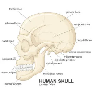
- Cranium: the brain-box.
- Facial skeleton: the bones of the face.
- Jaws: the maxilla (upper jaw) and the mandible (lower jaw).
What are the functions of the skull?
- It protects the brain.
- It protects the olfactory organ, the eyes, and the middle and inner ear.
- It gives shape to the head.
- It bears the teeth.
What is the vertebral column?
The vertebral column is the backbone, a series of bones called vertebrae that protects the spinal cord.

- Groups: the vertebrae are in five groups, each built on a similar basic pattern.
- Ligaments: strong ligaments hold the vertebrae together.
- Intervertebral discs: compressible cartilage pads between the vertebrae.
What are the types of vertebrae?
There are five types of vertebrae: cervical, thoracic, lumbar, sacral and caudal.
| Vertebrae | Location | Rat | Cat | Rabbit | Cow | Humans |
|---|---|---|---|---|---|---|
| Cervical | Neck | 7 | 7 | 7 | 7 | 7 |
| Thoracic | Chest | 13 | 12 to 13 | 13 | 12 | 12 |
| Lumbar | Lower back | 6 | 6 to 7 | 7 | 6 | 5 |
| Sacral | Hip region | 4 | 4 | 3 | 5 | 5 |
| Caudal | Tail | 30 | 16 | 18 to 25 | 18 to 20 | 4 |
What are the features of a typical vertebra?
A typical vertebra is a single bone of the backbone with the following parts.
- Neural canal: the passage for the spinal cord.
- Neural spine: projects upward and backward for the attachment of muscles.
- Transverse processes: for the attachment of muscles and ligaments.
- Centrum: a solid bony piece below the neural canal.
- Zygapophyses: surfaces that join successive vertebrae.
- Prezygapophyses: zygapophyses facing inward and upwards.
- Postzygapophyses: zygapophyses facing outward and downwards.
What are the cervical vertebrae?
The cervical vertebrae are the seven neck vertebrae, and the first two are the atlas and the axis.
The atlas (first cervical vertebra)
- It has a large neural canal.
- It has flat and broad transverse processes.
- It has a short neural spine, which may be absent.
- It has a vertebrarterial canal for the passage of blood vessels.
- The centrum is absent.
- Function: it permits the nodding of the head.
The axis (second cervical vertebra)
- It has a broad and flat centrum.
- It has a large and flat neural spine.
- It has reduced transverse processes and a vertebrarterial canal.
- It articulates with the atlas through the odontoid process.
- Function: it permits the turning or twisting of the head.
- Function: it forms a pivot joint with the atlas.
| Feature | Atlas | Axis |
|---|---|---|
| Position | First cervical vertebra | Second cervical vertebra |
| Centrum | Absent | Broad and flat |
| Neural spine | Short, may be absent | Large and flat |
| Transverse processes | Flat and broad | Reduced |
| Odontoid process | Absent | Present |
| Head movement | Nodding | Turning or twisting |
What are the thoracic vertebrae?
The thoracic vertebrae are the chest vertebrae that carry the ribs.
- They have a long and prominent neural spine.
- They have a pair of short transverse processes.
- They have a large neural canal and neural arch.
- They have a large cylindrical centrum.
- They have special surfaces for the attachment of the ribs.
Functions
- They aid the attachment of ribs.
- They assist in breathing.
- They give attachment to muscles at the shoulder and back.
What are the lumbar vertebrae?
The lumbar vertebrae are the strong vertebrae of the lower back.
- They have large and flat transverse processes.
- They have a broad and flat neural spine.
- They have a large and thick centrum.
- They have well-developed zygapophyses.
- They have extra paired projections: the anapophysis and the metapophysis.
Functions
- They provide attachment for abdominal muscles.
- They bear considerable weight of the body.
What are the sacral vertebrae?
The sacral vertebrae are the hip-region vertebrae that fuse into a single bony mass called the sacrum.
- Each has a narrow neural canal.
- Each has a reduced neural spine.
- Each has a large centrum.
The first sacral vertebra differs from the other four in two ways:
- It has a pair of large, wing-like transverse processes, while the others give attachment to the muscles of the back.
- It has a small neural canal, and the canal becomes narrower in the lower four vertebrae.
- Function: the sacrum joins the pelvic girdle to give support, rigidity and strength.
What are the caudal vertebrae?
The caudal vertebrae are the tail vertebrae, which are joined into a single bony mass called the coccyx in man.
- They have no neural spine, no neural canal and no transverse process.
- The coccyx appears as a solid rectangular mass of bone.
Functions
- It supports the tail.
- It provides attachment for tail muscle.
What are the ribs?
The ribs are long, semi-circular rods that connect the thoracic vertebrae to the sternum.

- Position: they lie in the chest region.
- Number: man has 12 pairs.
- Parts of a typical rib: a head, a neck and a body.
- Protection: they form a cage protecting the lungs and the heart.
- Breathing: they assist in breathing.
| Type of rib | Pairs | Attachment to the sternum |
|---|---|---|
| True ribs | 1st to 7th | Connected directly through costal cartilage |
| False ribs | 8th to 10th | Share a common attachment, each joined by costal cartilage to the rib above |
| Floating ribs | 11th and 12th | No connection to the sternum |
What is the appendicular skeleton?
The appendicular skeleton is the part of the skeleton made up of the limb girdles and the limbs.
What is the pectoral girdle?
The pectoral girdle is the girdle around the shoulder in man that supports the fore limbs.
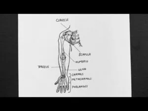
- Halves: it has two halves held by muscles.
- Scapula: flat and triangular, with a hollow called the glenoid cavity at its tip.
- Glenoid cavity: it articulates with the head of the humerus to form the shoulder joint.
- Coracoid: fixed to the scapula.
- Clavicle: a small rod of bone that connects the sternum to the scapula.
Functions
- It gives attachment to muscles and ligaments.
- It provides firm support to the fore limbs.
What is the pelvic girdle?
The pelvic girdle is the girdle around the waist in man that supports the hind limbs.

- Halves: two halves joined to each other ventrally and to the sacrum dorsally.
- Ilium: the first of the three bones in each half.
- Ischium: the second bone.
- Pubis: the third bone.
- Acetabulum: the depression formed on the outer surface by the three bones, which articulates with the head of the femur to form the hip joint.
What are the limbs?
The limbs are the fore (upper) and hind (lower) limbs, which share the same basic plan in most vertebrates.

- Basic plan: one long bone, then a pair of long bones, then a set of small bones ending in five digits.
| Region | Fore limb | Hind limb |
|---|---|---|
| Upper bone | Humerus | Femur |
| Pair of long bones | Radius and ulna (ulna is longer) | Tibia and fibula (tibia is longer and larger) |
| Joint between upper bone and pair | Elbow | Knee (with patella in front) |
| Small bones | Carpals (wrist) | Tarsals (ankle) |
| Digit bones | Metacarpals | Metatarsals |
| End of digits | Phalanges (finger bones) | Phalanges (toe bones) |
- Fore limb phalanges: in man, each finger has three phalanges except the thumb, which has two.
- Femur: the largest and longest bone in the body.
- Condyles: two rounded projections at the lower end of the femur, which form the knee joint together with the tibia.
- Patella: a small flat bone in front of the knee joint.
- Hind limb phalanges: in man, each toe has three phalanges except the big toe, which has two.
Evaluation
- Name the three forms of skeletal materials found in animals.
- State three differences between bone and cartilage.
- Name the three types of skeleton and give one example of an animal that has each.
- What is ossification?
- Name the two parts of the vertebrate skeleton and list the bones that make up each.
Assignment
Answer all the questions below in your exercise book.
- …………. is a non-living skeletal material. A. chondroblasts B. osteocyte C. elastic cartilage D. chitin
- The articulating surface for joining together successive vertebrae is called A. neural spine B. zygapophyses C. transverse processes D. neural canal
- The canal for the passage of blood vessels in vertebrae is called A. neural canal B. cervical canal C. vertebrarterial canal D. zygaphosis
- (a) What is ecdysis? (b) Mention two animals in which it occurs.
- Differentiate between (a) bone and cartilage (b) atlas and axis vertebrae.
This term's Biology topics
SS2 First Term · 9 topics
SS2 Biology Lesson Notes for Other Topics
Reproduction - Asexual Reproduction
Read the full lesson note.
Reproduction II - Sexual Reproduction
Read the full lesson note.
Reproductive Health
Read the full lesson note.
Cellular Respiration
Read the full lesson note.

Respiratory System
Read the full lesson note.
Excretory System
Read the full lesson note.

Supporting Tissues In Plants
Read the full lesson note.
Joints
Read the full lesson note.
Digestive System
Read the full lesson note.
Transport System
Read the full lesson note.
Circulatory System In Mammals
Read the full lesson note.
Mechanism Of Transportation In Higher Mammals
Read the full lesson note.
Cell Division
Read the full lesson note.
Reproductive System In Vertebrates
Read the full lesson note.
Reproductive System In Flowering Plants
Read the full lesson note.
Lesson Notes for Other Classes

SS1 Lesson Note
The complete lesson note to guide your studies.
SS3 Lesson Note
The complete lesson note to guide your studies.
