Circulatory System in Mammals – SS2 Biology Lesson Note (Second Term)
Download Lesson NoteLesson note at a glance
- Subject
- Biology
- Class
- SS2
- Term
- Second Term
- Topic
- Circulatory System in Mammals
- Order
- Topic 3 of 6 this term
- Reading time
- 6 min
The circulatory system of a mammal is made up of the blood, the heart and the blood vessels. The blood transports substances and protects the body against disease. Mammals have a closed, double circulation: blood stays inside vessels and passes through the heart twice in one complete trip around the body.
Behavioural objectives
By the end of the lesson, students should be able to:
- State the two basic functions of blood in mammals.
- Describe how the blood protects the body, including the mechanism of clotting.
- Distinguish between open and closed, and single and double, circulatory systems.
- List the types of blood vessel and state the differences between an artery and a vein.
- Describe the structure of the mammalian heart and the two phases of the heartbeat.
- Name the main arteries and the organs they supply.
What is the protective function of blood?
The blood performs two basic functions in mammals. These are:
- Transportation of various substances
- Protection of the body against diseases
The blood defends the body in the following ways:
- Antibody production (clumping): The lymphocytes produce antibodies. Antibodies are proteins normally present in the body or produced in response to an antigen, which they neutralise, thus producing an immune response. The same result can be achieved by injecting a small dose of weakened or dead pathogens into the body. This process is known as vaccination or immunisation.
- Neutralisation: The whole blood also produces antitoxins, which neutralise the toxins produced by the pathogens.
- Phagocyte action (engulfing): Phagocytes engulf the pathogens and digest them.
- Clotting: The blood platelets clot the blood to prevent germs from entering the body and to prevent loss of blood.
Mechanism of clotting
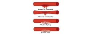
When a blood vessel is damaged and exposed to air, platelets in the bloodstream release an enzyme, thrombokinase. The thrombokinase converts prothrombin (an inactive blood protein) to an enzyme called thrombin. The thrombin then converts the soluble fibrinogen in the plasma to insoluble fibrin in the presence of calcium ions.
The threadlike fibrin forms a network or mesh on the surface of the wound, and blood cells are trapped within the mesh to form a clot. The clot dries to form a scab over the wound.
What are the types of circulatory system?
The circulatory system in animals can be:
- Closed or open
- Single or double
Closed circulatory system
Arteries divide into capillaries, which later join up with vessels called veins. Blood is therefore limited to the vessels and the heart, and it does not have direct contact with the body cells. This system is found in annelids and vertebrates.
Open circulatory system
The blood vessels lead out of the heart but end in blood spaces called haemocoels within the body cavity. The blood has direct contact with the cells and is then returned to the heart. Arthropods and some molluscs have an open circulatory system.
Single circulatory system
The blood passes through the heart once in one complete movement around the body. An example is the fish, whose two-chambered heart has a single circulatory system.
Double circulatory system
The blood passes through the heart twice every time it makes one complete movement around the body. Each time it passes through a separate pathway. Mammals are an example. Double circulation gives rise to pulmonary and systemic circulation.
What are the blood vessels?
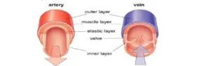
- Arteries: These are vessels that carry oxygenated blood away from the heart to the body’s organs, except for the pulmonary artery, which carries deoxygenated blood.
- Arteriole: A branch of an artery that gives rise to capillaries.
- Veins: These are large vessels that carry deoxygenated blood towards the heart, except for the pulmonary vein, which carries oxygenated blood.
- Venule: A small vessel that carries blood from the capillaries to the veins.
- Capillaries: They link the arteries with the veins around the tissues and organs. They are tiny and thin-walled to allow easy exchange of gases, nutrients and waste products between the cells and the blood.
Differences between an artery and a vein
| SN | Artery | Vein |
|---|---|---|
| 1 | It has a thick (muscular) wall | It has a thin wall |
| 2 | Elastic wall | Non-elastic wall |
| 3 | Carries blood away from the heart | Returns blood to the heart |
| 4 | Carries oxygenated blood, except the pulmonary artery | Carries deoxygenated blood, except the pulmonary vein |
| 5 | Blood is pink or bright in colour | Blood is dark red |
| 6 | Situated deep in muscles | Superficially situated |
| 7 | Has a small lumen | Has a large lumen |
| 8 | Pressure is high | Pressure is low |
| 9 | Pulse is readily detectable | Pulse is not easily detected |
| 10 | It has no valves, except semi-lunar valves | It has valves |
What is the heart?
The heart is a muscular and powerful organ responsible for pumping blood in the system of mammals. It is located within the chest cavity and is protected by the ribs and sternum. The pumping action of the heart is known as the heartbeat. The heart rate varies from animal to animal and often increases at moments of excitement. A human heart beats about 72 times per minute.
The special muscles that make up the heart are known as cardiac muscles. The heart is enclosed in a tough, two-layered protective membrane called the pericardium. Pericardial fluid fills the space between the heart and the pericardium. It reduces the friction arising from the pumping movement of the heart against its surrounding tissues.
What is the structure of the heart?
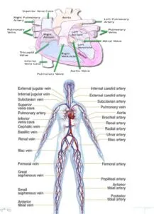
The human heart is divided into four chambers: the right and left auricles and the right and left ventricles. The walls of the ventricles are often thicker than those of the auricles. The left ventricle especially has a thick wall because it pumps blood out to all other parts of the body, and this requires more pressure.
The heart is divided into two halves by a central barrier called the septum. Bicuspid valves separate the left auricle and the left ventricle. This ensures that blood flows in one direction only, from the auricles to the ventricles. Similarly, the tricuspid valve exists between the right auricle and the right ventricle. It serves the same function as the bicuspid valve. These valves are held in place by special fibres and non-elastic cords known as chordae tendineae.
What is the heartbeat?
The heartbeat consists of alternate contraction and relaxation of the right and left auricles as well as the right and left ventricles. The human heartbeat is about 72 beats per minute. Each beat can be divided into two phases:
- Diastole: This is the first phase of the heartbeat. The two auricles contract, forcing blood into the ventricles, with oxygenated blood going into the left ventricle. As the ventricles fill up, the cuspid valves are pushed up and closed.
- Systole: This is the second phase of the heartbeat. The ventricles contract, sending blood out of the heart through the two main arteries. Deoxygenated blood from the right ventricle passes into the pulmonary artery, while oxygenated blood is sent into the aorta. The sequence then repeats itself.
Which organs do the main blood vessels supply?
| SN | Blood vessel | Organ supplied |
|---|---|---|
| 1 | Carotid artery | Head |
| 2 | Pulmonary artery | Lungs |
| 3 | Hepatic artery | Liver |
| 4 | Gastric artery | Stomach |
| 5 | Mesenteric artery | Intestine |
| 6 | Renal artery | Kidney |
| 7 | Gonadal artery | Gonads |
| 8 | Intercostal artery | Wall of thorax |
Corresponding veins accompany the arteries listed above.
Evaluation
- State the two basic functions of blood in mammals.
- Describe how blood clots when a blood vessel is damaged.
- Distinguish between an open and a closed circulatory system.
- State four differences between an artery and a vein.
- Name the valve between the left auricle and the left ventricle and state its function.
Assignment
- Name the organ supplied by each of these arteries: carotid, hepatic, renal and mesenteric.
- Explain why the wall of the left ventricle is thick.
- State the role of the pericardial fluid.
This term's Biology topics
SS2 Second Term · 6 topics
Second Term Biology Lesson Notes for Other Topics

Chromosomes
Read the full lesson note.
Variation
Read the full lesson note.
Adaptation
Read the full lesson note.
Evolution
Read the full lesson note.
Biological diagrams/drawing
Read the full lesson note.
Spelling Drills of Biological Technical Terms
Read the full lesson note.
Reproductive System In Vertebrates
Read the full lesson note.
Cell Division
Read the full lesson note.
Mechanism Of Transportation In Higher Mammals
Read the full lesson note.
Digestive System
Read the full lesson note.
Transport System
Read the full lesson note.

Relevance Of Biology To Agriculture
Read the full lesson note.

Energy Transformation In Nature
Read the full lesson note.

Functions In Ecosystem
Read the full lesson note.
Basic Ecological Systems III
Read the full lesson note.
Lesson Notes for Other Classes

Basic 1 Lesson Note
The complete lesson note to guide your studies.
Basic 2 Lesson Note
The complete lesson note to guide your studies.

Basic 3 Lesson Note
The complete lesson note to guide your studies.

Basic 4 Lesson Note
The complete lesson note to guide your studies.

Basic 5 Lesson Note
The complete lesson note to guide your studies.

Basic 6 Lesson Note
The complete lesson note to guide your studies.