Tissues And Supporting Systems SS1 Biology Lesson Note
Download Lesson NoteTopic: Tissues And Supporting Systems
INTRODUCTION
To carry out life processes, all organisms (plants and animals) need tissues. Tissues are a group of similar cells that carry out specific functions. The skeleton is the framework of the body which provides support, shape and protection to the soft tissues and organs in animals. It forms the central core of the human body and it is covered by muscles blood vessels and skin.
FORMS OF SKELETAL MATERIALS
There are 3 forms of skeletal materials found in animals. These are
- Chitin
- Cartilage
- Bones
CHITIN
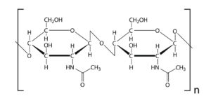
It is a tough non-living material present in arthropods (invertebrates). It acts as a hard outer covering for the animal and is made up of a series of plates covering or surrounding organisms. Chitin is very tough, light and flexible. However, it can be strengthened by impregnation with ‘tanned’ proteins and particularly in the aquatic crustaceans like crabs, by calcium carbonate.
CARTILAGE
This is a tissue present in the skeleton of complex vertebrates. Cartilage consists of a hard matrix penetrated by numerous connective tissue fibres. The matrix is secreted by living cells called chondroblasts. These later become enclosed in spaces (lacunae) scattered throughout the matrix. In this condition, the cells are termed chondrocytes. It acts as a shock absorber in between bones during movement because it is tough and flexible with great tensile strength. It is found predominantly in mammals and cartilaginous fishes e.g. sharks.
TYPES OF CARTILAGE
Cartilages are of three main types in mammals and they are:
i. HYALINE CARTILAGE
This contains dense meshwork is the most common type and can be found on the surface of moveable joints, trachea and bronchi (for ease of respiration) and also in protruding parts of the nose.
ii. WHITE FIBROUS CARTILAGE
Tougher than the hyaline cartilage and can be found in the intervertebral disc of the vertebral column.
iii. YELLOW ELASTIC CARTILAGE
Found in the external ear (pinna) and epiglottis (*cartilaginous flap covering the trachea active during food swallowing).
BONE
This is the major component of the skeletal system and it consists of living cells (osteocytes), protein fibres (collagen), and minerals such as calcium carbonate and calcium phosphate. These minerals (the non-living constituent) make up two-thirds of the mass of bone. Hence, bone is strong and very rigid, unlike cartilage. Bones are highly vascularized.
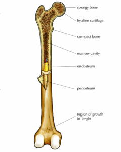
The skeleton of a young vertebrate embryo is made up of cartilage. As the embryo grows bone cells (osteocyte) replace cartilage cells. Hence, the cartilage tissue becomes hardened into bone through the addition of minerals in a process called OSSIFICATION
Differences Between Bones and Cartilage
| S/N | BONE | CARTILAGE |
| 1 | Bones produce red and white blood cells | Cartilages do not |
| 2 | Made up of both living cells and dead cells | Made up of mainly living cells. |
| 3 | Bones are often rigid | Cartilage is often flexible |
| 4 | They are made up mainly of mineral substances such as calcium | Mineral substances are absent |
| 5 | Can never be replaced by cartilage | Can be replaced by bones |
| 6 | Flexible only in young ones | Cartilage both in young ones and adults is flexible. |
TYPES OF SKELETON
The three main types of skeletons in animals are:
- Hydrostatic skeleton: This is the type present in soft-bodied animals e.g. earthworms, sea anemones etc. Such animals use pressure to support themselves. They also have a muscular body wall which is filled with fluid. The fluid presses against the muscular wall causing them to contract and exert force against the fluid.
- Exoskeleton: This is the outer skeleton present in arthropods. It is secreted by the cells covering the body of the animals and the main component is chitin (a non-living substance). The exoskeleton also supports animals against gravity and enables them to move about. Animals with these skeleton types periodically shed the old skeleton, and grow rapidly in size when the new exoskeleton is still soft and extensible. The shedding process is called MOULTING or ECDYSIS.
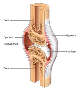
- Endoskeleton: This is an internal skeleton present in all vertebrates. The Endoskeleton of vertebrates are composed mainly of bones and the bones grow steadily as the animal grows (hence no need for moulting). Bones of many sizes and shapes make up the endoskeleton of vertebrates. These bones are attached as moveable joints by tough flexible fibers called ligaments hence the skeleton is flexible. Muscles are also attached to the bones usually by tendons to provide posture and bring about body movement.
FUNCTION OF SKELETON
- It supports the body of organisms.
- Skeleton acts as the framework of the body
- Protection of delicate organs e.g. heart, brain, etc.
- Used for locomotion through the limbs in action.
- Important components of respiration e.g. breathing involve active movement of the ribs.
- Production of blood via bone marrow.
SUPPORT IN VERTEBRATES
The skeleton of vertebrates such as fish, frogs, lizards, birds and man consists of bones and cartilages. It can be classified into two.
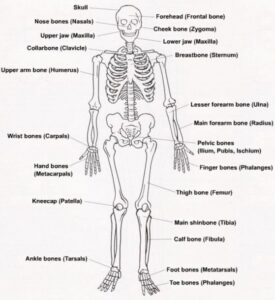
- AXIAL SKELETON: which consists of the skull, ribs, sternum and the vertebral column.
- APPENDICULAR SKELETON: it is made up of limb girdles (pectoral and pelvic girdles), and the limbs (the fore and hind limbs).
Types of Vertebrae and Location
| VERTEBRATE | LOCATION | RAT | RABBIT | CAT | COW | HUMANS |
| Cervical | Neck | 7 | 7 | 7 | 7 | 7 |
| Thoracic | Chest | 13 | 12 – 13 | 13 | 13 | 12 |
| Lumber | Upper trunk | 6 | 6 – 7 | 7 | 6 | 5 |
| Sacral | Lower trunk | 4 | 4 | 3 | 5 | 5 |
| Caudal | Tail | 30 | 16 | 18 – 25 | 18 – 20 | 4 |
-
AXIAL SKELETON
a) The Skull
The Skull is made up of flat bones joined together by a suture joint which has three parts: Cranium (brain-box), facial skeleton and the jaws; including the maxilla (upper) and mandible.
Functions
- It protects the brain.
- Also protects the olfactory organ, eyes, middle and inner ear.
- Gives shape to the head.
- Bears the teeth.
b) The vertebral column
It forms the backbone, protecting the spinal cord. It is made up of 5 groups of bones called the vertebrate each of which is built on a similar basic pattern. The vertebrate is held together with strong ligaments and comprehensible cartilage pads called into the intervertebral disc.
A typical vertebra has the following structural features:
– Neural canal which is for the passage of the spinal cord.
– Neural spine which projects upward and backward for the attachment of muscle.
– Transverse processes for the attachment of muscles and ligaments.
– Centrum; a solid bony piece below the neural canal
– Zygapophyses are the particular surfaces for joining together of successive vertebrates. This could be pre-zygapophysis (facing inward and upwards) or post-zygapophysis (facing outward and downwards
c) Cervical Vertebrae
The first cervical vertebra is called the atlas while the second is called the axis.
- The Atlas: It has a large neural canal, flat and broad transverse processes, short neural spine which could be absent at times. It also has a vertebraterial canal for the passage of blood vessels. Centrum is absent.
The function of Atlas
i. Permits the nodding of the head.
2.The Axis: It has a broad and flat Centrum, a large and flat neural spine, reduced transverse processes a vertebrarteria lcanal. It articulates with the atlas through the odontoid process
Functions
– It permits the turning or twisting of the head.
– Forms pivot joint with the atlas.
d) Thoracic Vertebra
Have a long and prominent neural spine, a pair of short transverse processes, a large neural canal and neural arc and a large cylindrical centrum. They also have particular surfaces for attachment of the ribs.
Function
- Aids attachment of ribs
- Assist in breathing
- Attachment of muscles at the shoulder and bac
e) Lumbar Vertebrae
Each has large and flat transverse processes, broad and flat neural spine, large and thick centrums and well-developed zygapophyses. It has extra paired projections namely
- Anapophysis
- Metapophysis
Functions of Lumbar
– It provides attachment for abdominal muscles
– It bears considerable weight on the body
f) Sacral Vertebrae
This fuse together to form a singular bony mass called the sacrum. Each sacral vertebrate has a narrow neural canal, reduced neural spine and large centrums. The first differs from the remaining four by having a pair of transverse processes which is large and wing-like while the others are attached to the muscles of the back.
The presence of a small neural canal generally becomes narrower in the lower four vertebrae.
FUNCTION
– Joins the pelvic girdle to provide support, rigidity and strength.
g) Caudal vertebrae
These are joined together to form a singular bony mass called a coccyx. Each has no neural spine, no neural canal and no transverse process. It appears as a solid rectangular mass of bone
Functions
- Supports the tail
- Provides attachment for tail muscle
ii. The Appendicular Skeleton
a) Pectoral girdle: found around the shoulder in man and it consists of two halves which are held by muscles. Each halve is made up of 3 three bones
i. Scapula
ii. Clavicle
iii. Coracoids
I. The scapula and coracoids are fixed together as the scapula is flat and triangular with a hollow called GLENOID CAVITY at its tip. This cavity articulates or joins with the head of the humerus to form the shoulder joint. The clavicle is a small rod of bone attached to a ligament joining the sternum to the scapula
Functions
a) The pectoral girdle gives attachment to muscles and ligaments.
b) It provides firm support to the forelimbs.
b) Pelvic girdle: found around the waist in man and it consists of two halves which are joined to each other ventrally and to the sacrum dorsally. Each half of the pelvic girdle is made up of 3threebones. They are:
a) Illium
b) Ischium
c) Pubis
These three bones form a depression (on their outer surface) called ACETABULUM which articulates with the head of the femur to form the hip joint.
c) LIMBS
The limbs include the fore (upper) and the hind (lower) limbs. In most vertebrates, both limbs have the same basic plan i.e. each limb has a long bone followed by a pair of two long bones next to this is a set of small bones terminating with five digits.
- The forelimbs- This is made up of an upper arm bone called the humerus which joins with two other long bones at its lower end (radius and ulna) to form the elbow joints. Radius and ulna (the ulna is longer) are the bones of the forearm, next are the wrist bones called carpals which are small bones. These are followed by the digit bones called metacarpals which terminate in the phalanges (finger bones). In man, each digit has three phalanges except the thumb which has two phalanges.
- The hind limbs are made up of thigh bones called the femur (which is the largest and longest bone in the body). Its round upper end is the end that terminates at two rounded projections called condyles which form the knee joint together with the tibia. A small flat bone called patella is found in front of the knee joint. Next to the femur are the tibia and fibula-Tibia is longer and larger. These are followed by bones of the ankle called tarsals. The lower limb terminates at the digit bone metatarsals and each digit is made up of three phalanges
- The ribs: These are long semi-circular rods which connect the thoracic vertebrates to the sternum. They are found in the chest region of the body. In man, there are 12 pairs
Function
– They form a cage protecting the lungs and the heart
– They assist in breathing.
A typical rib has a head, a neck and a body. The first seven ribs are connected directly to the sternum through costal cartilage. They are therefore called true ribs. The next five are called false ribs. The eighth to tenth ribs have a common articulation to the sternum, each one attached to the costal cartilage to the one above. The eleventh and twelfth pairs of ribs are called floating ribs because they have no connection to the sternum.
SUPPORTING TISSUES IN PLANTS
The needs for supporting tissues in plants are for:
- Definite shape;
- Strength;
- Rigidity;
- resistance against external forces such as wind and water.
Types of Supporting Tissues
- Parenchyma tissues
- Collenchyma tissues
- Sclerenchyma tissues
- Xylem tissues
- Phloem tissues
1.Parenchyma Tissues
They are made up of living cells with cellulose and many air spaces between them. This is the most common and abundant plant tissue.
Functions
- It gives firmness and turgidity to the stems of hibiscus
- Stores food and water
iii. Takes part in food synthesis in leaf mesophyll
2.Collenchyma Tissues
Made up of living cells which are elongated and thickened at the corners.
Functions
- Provides strength and support in young growing plants.
- Gives flexibility and resilience to plant.
3.Schlerenchyma Tissues
They are made up of thick cells containing cellulose and lignin. The tissues are rich in fibres.
Functions
- Gives flexibility to plant
- Provides strength, rigidity, hardness and support to plant
4. Xylem
Xylem tissues are found in vascular tissues of stems, roots and leaves
Functions
- Provides support strength and shape to the plant
- Helps to conduct water and mineral salt from the roots to the leaves.
5.Phloem Tissues
Also located in the vascular bundles of all plants in their roots, stems and leaves
Functions
- Conduction of manufactured food from the site of production to the site of consumption and storage.
- Assist in providing support to the entire plant.
ASSIGNMENT
- …………. is a non-living skeletal material
A. chondroblasts B. osteocyte C. elastic cartilage D. chitin
2. The articulating surface for joining together successive vertebrates is called A. neural spine B. zygapophyses C. transverse processes D. neural canal
3. The canal for the passage of blood vessels in vertebrae is called A. neural canal B. cervical canal C. vertebrate rial canal D. kyphosis
4. Endo-skeleton is present in the following animals except A. dog B. snake C. shark D. lizard
5. The most abundant supporting tissue in plants is A. sclerenchyma B. parenchyma C. xylem D. phloem
6(a) What is ecdysis?
(b) Mention two animals in which it occurs
7Differentiate between
(a) Bone and cartilage
(b) Atlas and axis vertebrae

Second Term Biology Lesson Notes for Other Topics
Chromosomes
Explore lesson notes covering all topics.
Variation
Explore lesson notes covering all topics.
Adaptation
Explore lesson notes covering all topics.

Evolution
Explore lesson notes covering all topics.
Biological diagrams/drawing
Explore lesson notes covering all topics.
Spelling Drills of Biological Technical Terms
Explore lesson notes covering all topics.
Reproductive System In Vertebrates
Explore lesson notes covering all topics.
Cell Division
Explore lesson notes covering all topics.
Mechanism Of Transportation In Higher Mammals
Explore lesson notes covering all topics.

Digestive System
Explore lesson notes covering all topics.
Circulatory System In Mammals
Explore lesson notes covering all topics.

Transport System
Explore lesson notes covering all topics.
Relevance Of Biology To Agriculture
Explore lesson notes covering all topics.
Energy Transformation In Nature
Explore lesson notes covering all topics.
Functions In Ecosystem
Explore lesson notes covering all topics.


Lesson Notes for Other Classes
JSS1 Lesson Note
The complete lesson note to guide your studies.
JSS2 Lesson Note
The complete lesson note to guide your studies.

SS2 Lesson Note
The complete lesson note to guide your studies.

SS3 Lesson Note
The complete lesson note to guide your studies.